MRI Scans Explained
What is an MRI scan?
MRI scanning is a powerful medical diagnostic tool that uses a strong magnet to produce high-quality images in different planes. Magnetic resonance imaging uses non-ionising radiation for imaging, unlike x-rays which uses x-ray radiation.
An MRI scanner consists of a strong magnet with a radio transmitter and receiver. These instruments gather the information out of your body. MRI produces soft-tissue images and is used to distinguish normal, healthy soft tissue from pathologic tissue.
The human body is largely made of water molecules with hydrogen and oxygen atoms. At the center of each atom lies an even smaller particle caled a proton, which is sensitive to any magnetic field. Normally the water molecules in the body are randomly aligned, but upon entering an MRI scanner, the magnetic field causes the body's water molecules to align uniformly in one direction. The gradient amplifiers then deliver a series of short RF pulses, which causes the hydrogen atoms to alter their alignment. When the RF pulse is switched off, the hydrogen atoms relax back to their original state and in the process give out a weak signal. The patient cannot feel these changes, but the sensitive coils placed on the patient, can pick up these signals. These are amplified and then processed by special computer-based image processing algorithms to create detailed sectional images of the body for the Radiologist to interpret.
Why would I need an MRI scan?
An MRI scan is one of the most sophisticated diagnostic tools available to help a referring clinician understand the cause of your particular health issue.
What can be diagnosed by an MRI scan?
By scanning the relevant parts of a patient's body, an MRI scan can help to diagnose a patient's medical problems like:
- ailments of the brain, including tumours and dementias
- sports injuries
- musculoskeletal problems
- most spinal conditions/injuries
- vascular abnormalities
- female pelvic problems
- prostate problems
- abdomen and gastrointestinal tract conditions
- certain ear, nose and throat (ENT) conditions
- soft tissue and bone pathology/conditions
- and other clinical conditions as may be decided by the clinician
Who can't have an MRI scan?
MRI is a non-invasive and safe test. As MRI works with a strong magnet and radio waves, you need to tell us, if anything of the following applies to you or the person that accompanies you into the MRI room.
You must not have an MRI scan if you have:
- a cardiac (heart) pacemaker
- certain clips in your head from brain operations, i.e. aneurysm clips
- a cochlear (ear) implant
- had surgery in the last 8 weeks
- a programmable shunt for hydrocephalus (fluid on the brain)
- Implanted cardioverter defibrillator (ICD)
- Electronic implant or device
- Magnetically-activated implant or device
- Neurostimulation system?or Spinal cord stimulator
- Insulin or infusion pump?or Implanted drug infusion device
- Any type of prosthesis or implants
- Artificial or prosthetic limb
- Any external or internal metallic object?in your eye or body
- if you are pregnant
- However the individual cases could be discussed with our Radiologist and a final decision taken.
Preparing for an MRI Scan
Carry all your previous reports and studies along with you.
No special preparation is needed prior to the exam, unless your doctor has given you other instructions.?You will be asked to complete a safety screening form and answer questions pertaining to your medical history.
Before your scan, you will be asked to change into loose clothing without zippers or metallic parts, which will be provided by our staff. Also you will be asked to remove all metallic items such as jewelry?, watches, hairpins?, glasses?, wallets? and other metallic objects, which can be stored in lockers or left with your attenders, prior to the scan.
Do I need contrast for the MRI scan?
Depending on the clinical information your doctor needs, the MRI scan may require the use of a contrast-agent given intravenously to assist in visualization of certain structures in your body to aid the diagnosis. This will be decided by your clinician or our Radiologist prior to or during your study. Details regarding the contrast media and any possible side effects, though minimal, will be explained by our Radiologist prior to contrast administration.
Preparations for Contrast study
The patient should come for examination with minimum 3 hrs of fasting. However patients who cannot tolerate long hours of fasting, especially diabetics can take watery fluids, after discussion with our staff. You will be asked to take a S Creatinine blood test prior to the scan and bring the results along with you. Special precautions are needed for Diabetic patients and those with a history of allergic conditions. Please discuss with our staff regarding these prior to your scan.
What happens during an MRI Scan?
After you have changed and removed all metal objects, the technologist will position you onto the MRI table, place a small coil on you and the table will then slide into the scanner. Our MRI units feet-first functionality allows that your head stays outside the magnet bore during most examinations (with the exception of head/neck/chest scans), which is very comfortable for patients. Anyway you will be able to communicate with the technologist during the scan through the intercom. You will also be given an emergency call button to call our staff in the event of any issue
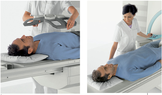
For clear pictures, you will be asked to hold very still and relax. In some cases, you will be asked to hold your breath. Any movement, especially of your head or back (even moving your jaw to talk) during the scan will blur the pictures. While the machine is taking your pictures, you will hear rapidly repeating, thumping noises coming from the walls of the scanner. For additional comfort, earplugs may be provided. During this time, you could breath quietly and normally, but otherwise refrain from any movement, coughing or wiggling. When the thumping noise stops, you must refrain from changing your position or moving about.
This whole procedure will usually be repeated several times, and?the entire exam ordinarily takes between 15 and 30 minutes to complete.